


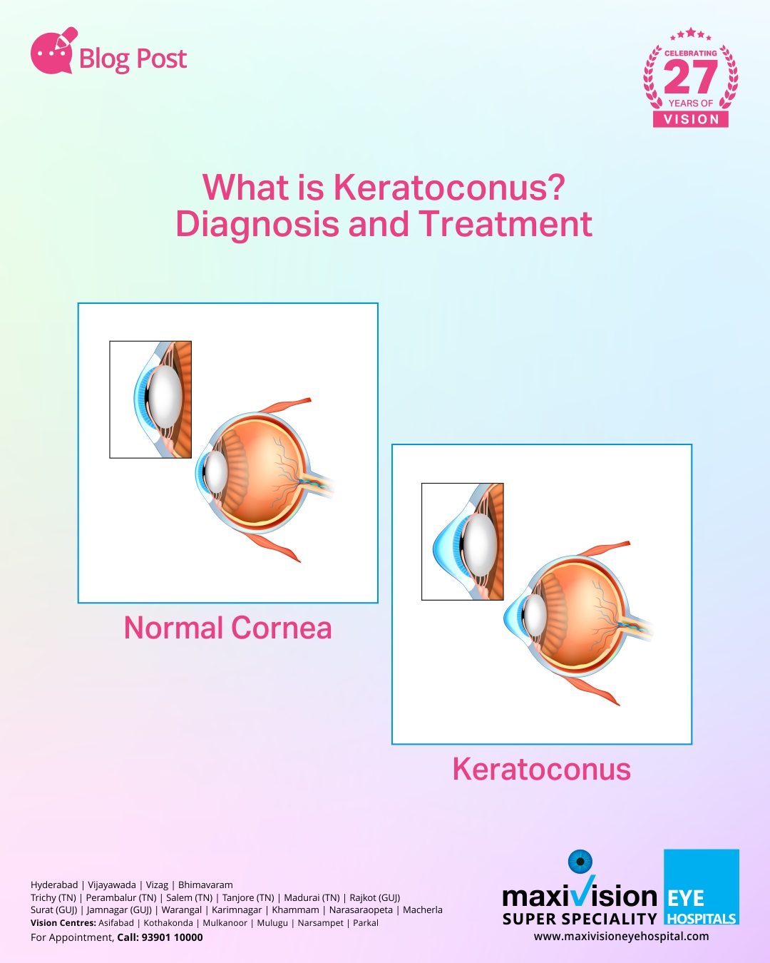
Keratoconus refers to the bulging of the eyes in the shape of a cone. This happens when the collagen structure of your cornea weakens and cannot hold the regular round corneal shape in place. It’s usually seen in the 20s but can also start in childhood.
In old age, Keratoconus is observed when it’s mild. There’s no clear explanation about its causes except that it could be hereditary or due to certain medical conditions.
Keratoconus affects vision in two ways.
- It causes irregular astigmatism, which can’t be fully corrected with glasses due to the warping of the corneal surface.
- As the cornea steepens slowly, the vision changes and hence, you become near-sighted. This may require frequent changes of glasses to accommodate the power change.
Symptoms of Keratoconus
Usually, people with Keratoconus will observe vision distortion slowly. This may end at any time or continue for years.
The main symptoms include
- You will notice halos around bright lights
- Eyesight gradually gets worse towards the end of adolescence
- You could experience double vision when looking with only one eye, even when you wear spectacles
- Light streaks
- Triple ghost images
- Blurry vision
Diagnosis of Keratoconus
Your eye doctor will review your family history apart from conducting an eye exam. The main tests done include
- Keratometry: A circle of light from a device falls on your cornea to measure the reflection of light. Thus, it helps determine the corneal shape.
- Slit-lamp examination: Here, a vertical light beam projection happens on your eye, and a low-power microscope helps view the eye for corneal shape abnormalities and other potential problems.
- Eye refraction: This device measures your eyes using wheels of lenses. Hence, this device helps determine your sharpest vision using a multiple-lens combination.
- Computerised corneal mapping: The main tests done are tomography and topography. These tests create a detailed map of your cornea, thus enabling your doctor to check for signs of Keratoconus.
Tomography also helps measure the thickness of your cornea.
Treatment of Keratoconus
The severity of the condition determines the treatment method. Two approaches to treating the conditions involves
- Slowing the disease progress
- Improving vision
Corneal Collagen Crosslinking, or C3R, is the treatment method for worsening conditions. Your cornea is saturated with riboflavin drops and treated with UV light. Hence, it strengthens your cornea and allows the stabilising of power using glasses. You may not need a cornea transplant in such a scenario.
Different lenses used in the treatment of Keratoconus
- Soft contact lenses: You can use them in early Keratoconus, but they’ll have to change with your increasing eye bulge.
- Hard contact lenses, including rigid and gas-permeable lenses, treat advanced Keratoconus.
- Other lenses include piggyback, hybrid and scleral lenses.
Conditions such as corneal scarring, extreme thinning of your cornea, poor vision or inability to wear any contact lenses will require you to undergo surgery. This can be a corneal transplant or intrastromal corneal ring segments (ICRS) application.
Hence, you can keep Keratoconus in check through proper detection and treatment. Your alertness matters the most.
Maxivision is the best eye hospital in Hyderabad. Hence, if you have symptoms of Keratoconus, please consult our expert eye specialists for the best care.


+ There are no comments
Add yours